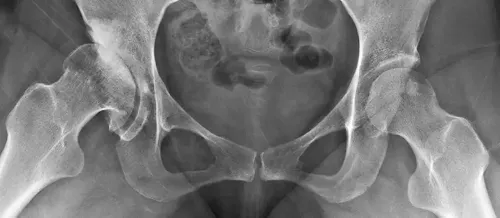
Dysmetries and shortening of long bones

What is dysmetria?
Bone dysmetry is the discrepancy in the length of the extremities, either by excess (hypermetry) or by default (hypometry). It may be congenital or acquired due to fractures or infections in the leg bones.
Limb dysmetry is a frequent reason for consultation in children's orthopedics.
The longitudinal growth of the bone is related to the growth cartilages (physis). Each physis has its own potential for growth and in relation to bone age.
In the Department of Orthopedic Surgery and Traumatology of the Clínica Universidad de Navarra we have a specialized consultation in Bone Lengthening and correction of Complex Deformities with professionals with great experience in the therapeutic approach of this type of problems.

What are the symptoms of dysmetry?
The main clinic of lower limb discrepancies results in a gait disturbance, in addition to the aesthetic repercussion.
In these patients, an irregular and unstable gait is produced.
The compensation of an unstable gait is produced by the inclination of the pelvis towards the side of the short extremity and deviation of the spine in the opposite direction.
On the other hand, it is generally accepted that dysmetries greater than 2.5 cm. in adulthood can produce low back pain and scoliotic attitude.
The most common symptoms are:
- Gait disturbance.
- Limping when walking or running.
- Discomfort when walking.
- Pain in the legs or hip or knee joints.
- Scoliotic posture (deviated to one side).
Do you have any of these symptoms?
You may have a dysmetry
What are the causes of dysmetria?
Dysmetries can have various causes, both congenital and acquired.
Congenital causes refer to those that are present from birth, with fibular hemimelia and congenital short femur being the most common malformations requiring reconstructive techniques.
Acquired causes are those that develop later in life due to growth plate injuries, complex fractures or infections in the leg bones (osteomyelitis).
Some possible causes of dysmetria include:
- Congenital bone or joint malformations.
- Traumatic injuries, such as fractures.
- Bone or joint infections.
- Bone growth disorders.
- Neuromuscular diseases that affect limb length.
Complications of dysmetria
Differences in limb length can lead to a number of complications that affect both physically and emotionally. Some of the most common complications include:
- Chronic pain: People with dysmetria may experience chronic pain in the joints and muscles of the legs due to uneven loading and difficulty walking properly.
- Deformities: In severe cases of untreated dysmetria, bony deformities may develop in the spine, hips and knees as a result of the compensation made by the body to adapt to the difference in leg length.
- Functional limitations: Dysmetria can limit a person's ability to carry out normal daily activities, such as walking, running or even standing for long periods of time.
- Postural problems: Lack of proper alignment can negatively affect an individual's posture, which can lead to back pain, neck and shoulder problems, and breathing difficulties.
- Joint wear and tear: Uneven loading on the joints can lead to premature wear and tear, which increases the risk of developing arthritis in the future.
How is dysmetry diagnosed?

In congenital causes, the diagnosis is usually early by intrauterine manifestations assessed by ultrasound methods. It is recommended to perform the general examination with the patient in underwear, in order to assess skin aspects, length of upper limbs, facial symmetry, etc.
At early ages (especially the first year of life), the examination of the hip is of vital importance for the evaluation of dysplastic phenomena.
For this reason, it is advisable to have a simple radiological examination in the anteroposterior projection of the pelvis.
The CT scan is the most accurate method, since it allows the assessment of the axes, without magnification. Based on these techniques, and especially through conventional radiology, limb alignment exams can be performed.
These studies will allow us to define the type of deformity, locate and measure as accurately as possible the axial deviations of each segment and compare them with ranges of normality. In addition, they will allow us to measure the dysmetry radiologically, as well as to plan possible correction methods.
How is dysmetry treated?
Choice of treatment according to the type of dysmetria
Usually the type of treatment is chosen according to the magnitude of the discrepancy.
Dysmetries of less than 1 cm are usually well tolerated and only require periodic controls in stages of growth.
- Differences between 1-3 cm. are tributary of compensatory rises.
- Dysmetries greater than 3 cm. are usually treated with surgical methods: Patients with a prognosis of dysmetry between 3 and 7 cm. can be treated with epiphysiodesis, or with lengthening techniques, while those predicted to have dysmetry greater than 7 cm. are usually treated by lengthening, in one or more surgical times.
- In cases of severe deformities, with a prognosis of severe dysmetry, amputation should be considered as a valid option for rapid adaptation of the patient to the prosthetic material.
There are two fundamental options for limb dysmetry:
Bone shortening of the long limb
The shortening techniques, by bone resection, have the disadvantage of operating on a healthy limb and reducing the patient's size.
It is also possible to perform growth plate blocks in children, which have the advantage of being less complex surgeries but, on the other hand, they are not very accurate. Usually these types of bone shortening are indicated in moderate dysmetries of 3-4 cm.
Bone lengthening
Currently, due to technical advances in recent years, bone lengthening techniques are the most commonly used to treat dysmetria.
External fixators
Conventional lengthening techniques, still used in some centers, consist of making a transverse cut in the bone and applying progressive distraction on both sides of the cut (osteotomy) to achieve progressive separation of the fragments and thus increase the length of the bone at a rate of approximately 1mm/day.
External fixators are used to achieve this distraction. There are many types of this device but they have the common denominator of being anchored to the bone from the outside by means of screws or pins and having a distraction mechanism that is operated from the outside.
Although they have had their usefulness, these techniques have many disadvantages due to the connection with the outside for a long time, which can cause infections, pain or joint stiffness.
Lengthening with intramedullary nails
The two techniques most commonly used today are: lengthening on nail, which combines external fixators and conventional intramedullary nails, reducing by more than 60% the time needed to wear an external fixator; the other technique is lengthening with expandable (telescopic) endomedullary nails, which do not require any type of external fixator.
These telescopic nails have inside them a magnetic or electric mechanism that, activated by external remote control, makes them expand progressively, achieving a gradual increase in the length of the bone.
This surgery is performed with minimally invasive techniques so it is much better tolerated, avoid infections and improve joint recovery, reducing the tendency to infections.
All these techniques are complex and can present complications, it is important to put yourself in the hands of a specialized team with experience and the patient should be informed of the entire process.
The Department of Orthopedic Surgery and Traumatology
of the Clínica Universidad de Navarra
The Department of Orthopedic Surgery and Traumatology covers the full spectrum of congenital or acquired conditions of the musculoskeletal system including trauma and its aftermath.
Since 1986, the Clinica Universidad de Navarra has had an excellent bank of osteotendinous tissue for bone grafting and offers the best therapeutic alternatives.
Organized in care units
- Hip and knee.
- Spine.
- Upper extremity.
- Pediatric orthopedics.
- Ankle and foot.
- Musculoskeletal tumors.

Why at the Clinica?
- Experts in arthroscopic surgery.
- Highly qualified professionals who perform pioneering techniques to solve traumatological injuries.
- One of the centers with the most experience in bone tumors.
Our team of professionals
Specialists in Orthopedic Surgery and Traumatology with experience in treating dysmetria
Frequently asked questions about dysmetria
In some mild cases of dysmetria, there may be spontaneous improvement without treatment. This may occur especially during growth, as differences in limb length may gradually balance out.
However, it is important to note that this is not always the case and in more severe or progressive cases the dysmetria is unlikely to improve on its own.
For proper evaluation and treatment recommendations, it is essential to consult a specialist in Traumatology.
The ideal age for surgery in cases of dysmetria can vary with each individual and is determined by the severity of the leg length discrepancy, the patient's age and other factors.
In general, bone lengthening surgery is performed when the patient is considered to have reached sufficient skeletal maturity, which usually occurs in adolescence.
However, it is important to note that the optimal time for intervention should be determined by a specialist based on the evaluation of the specific case.
While bone lengthening surgery is a commonly used option to correct the difference in leg length, there are other non-surgical alternatives that can be considered in less severe cases.
One such option is the use of shoe lifts, which involves adding additional elevation to the sole of the shoe on the side of the shorter leg. This helps to balance the height and improve function during gait.
However, it is essential that any treatment decision be discussed and agreed upon with a specialized medical professional, who will assess the severity of the dysmetria and recommend the best option for each case.


