Femoroacetabular shock
"Through minimally invasive surgery we are able to correct the defects of the hip, achieving very good results".
DR. PABLO DÍAZ DE RADA LORENTE
SPECIALIST. ORTHOPEDIC SURGERY AND TRAUMATOLOGY DEPARTMENT

Femoroacetabular shock is a pathology of the hip joint, the origin of which is currently unknown, which can affect either of the two elements involved in hip play: the acetabulum and the femur.
The acetabulum is the concave articular portion of the surface of the pelvis, with which the head of the femur is articulated, forming the hip joint.
It mainly affects young people who practice sports. The lack of sphericity of the head of the femur causes pain when performing certain activities.

What are the symptoms of femoroacetabular shock?
Empieza a manifestarse en la mayoría de los casos como un dolor inguinal y, con menor frecuencia, en la región trocanterea (cadera), glúteos o en la cresta ilíaca.
El dolor se presenta durante o después de la práctica deportiva, cuando se permanece un rato sentado o bien al levantarse, después de estar un tiempo sedente.
También puede manifestarse como una lenta pérdida de la movilidad del juego de la cadera, a veces sin dolor alguno.
The most common symptoms are:
- Groin pain.
- Functional disability.
- Discomfort when walking.
Do you have any of these symptoms?
You may have femoroacetabular shock
What are the causes of femoroacetabular shock?
Femoroacetabular shock is usually due to the presence of a bulge in the neck of the femur (CAM) or in the acetabulum (PINCER).
1. CAM-type shock usually occurs in young, active, male patients. The femoral neck bulge collides with the acetabular rim and will result in injury to the adjacent cartilage in the acetabulum.
2. The anterolateral edge of the acetabulum protrudes so much that it collides with the neck of the femur in flexion gestures and hip rotations (Pincer impingement). It occurs more frequently in middle-aged athletic women
It is not known for sure why imperfect growth of affected bones occurs.
Trauma to the hip, or sports that cause repeated friction of the femur against the pelvis, with forced and maintained squatting positions (such as those of ice hockey players, or motorcycle riders), may result in labral (cartilage ring surrounding the acetabulum) injuries.
How is femoroacetabular shock diagnosed?

Various studies have shown that femoroacetabular shock, which is suspected of affecting 15% of the population, could be the cause of up to 70% of the arthroses hitherto considered idiopathic, which affect people under 50 years of age and which can end up in prostheses.
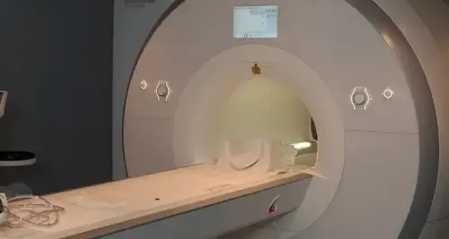
The personal interview with the patient, in addition to the diagnostic tests (radiography, magnetic resonance) are fundamental to confirm the pathology.
X-rays reveal bone imperfections and MRI helps us visualize possible injuries.
How is femoroacetabular shock treated?
Once the problem has been diagnosed, the first proposals to the patient are the use of anti-inflammatory medication and a physical therapy protocol that helps correct the harmful movements, while relieving the pain.
The intra-articular infiltrations usually reduce or make the pain disappear, sometimes temporarily and other times for a long period.
They fulfill two functions, on the one hand if the pain comes from the hip (and not of other near zones as it could be the spine, the pubis, the buttocks) the medication infiltrated in the interior will alleviate the pain.
At the same time, the diagnosis is confirmed. If the pain is relieved, it is another sign that the pain is coming from the hip and not the spine.
Depending on the degree of degenerative joint deterioration and the start of the clinic, there are the following therapeutic options:
- Hip arthroscopy, for the repair of grade 1 injuries.
- Femoroacetabular Osteoplasty. With this procedure, an enlarged circumduction is achieved, which eliminates the femoroacetabular shock. The authors indicate this technique in grades 1 and 2.
- Corrective osteotomies, which provide a more congruent articulation. All osteotomies will also be reserved for grades 1 and 2.
- Arthroplasty or replacement by a hip prosthesis. Grade 3.
Where do we treat it?
IIN NAVARRE AND MADRID
The Department of Orthopedic Surgery and Traumatology
of the Clínica Universidad de Navarra
The Department of Orthopedic Surgery and Traumatology covers the full spectrum of congenital or acquired conditions of the musculoskeletal system including trauma and its aftermath.
Since 1986, the Clinica Universidad de Navarra has had an excellent bank of osteotendinous tissue for bone grafting and offers the best therapeutic alternatives.
Organized in care units
- Hip and knee.
- Spine.
- Upper extremity.
- Pediatric orthopedics.
- Ankle and foot.
- Musculoskeletal tumors.

Why at the Clinica?
- Experts in arthroscopic surgery.
- Highly qualified professionals who perform pioneering techniques to solve traumatological injuries.
- One of the centers with the most experience in bone tumors.


