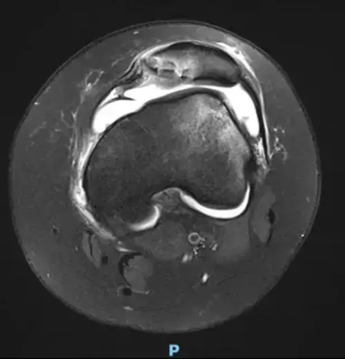
Meniscus injuries. Broken meniscus
"In patients who have lost a very important part of the meniscus and are young in age, replacement and treatment by means of a meniscal transplant may be recommended".
DR. ANDRÉS VALENTÍ
SPECIALIST. ORTHOPEDIC SURGERY AND TRAUMATOLOGY DEPARTMENT

The menisci are two fibrocartilaginous structures located in the knee joint between the femur and the tibia, one internal or medial and the other external or lateral.
They have a crescent shape and their function is to cushion and stabilize the knee joint allowing a better distribution of the load.
The menisci are important for the stability and functionality of the knee joint, and they also absorb shocks and decrease cartilage wear.
When they are broken they produce pain in greater or smaller lateral and posterior measurement, the knee can be blocked total or partially, with limitation in torsion gestures, forced flexion among others...

What are the symptoms when the meniscus is damaged?
The meniscal injuries in which a break of the meniscus occurs, generally, they go with medial or lateral pain to tip of finger depending on that the broken meniscus is internal or external and sometimes it produces pain in zone later of the knee.
The most common symptoms are:
- Pain and stiffness
- Inflammation and joint effusion
- You can study with a blocking
Do you have any of these symptoms?
You may have suffered a torn meniscus
What are the causes?
The breakage of a meniscus can occur for various reasons.
Traumatic: In young and active people, it is frequent its breakage by means of a torsional mechanism, sudden turn or traumatism.
Degenerative: As the knee ages, the meniscal structure loses elasticity and is subjected to more stress due to degenerative changes in the cartilage and progressive wear of the knee. The meniscus can break in everyday situations, without a clear history that the patient remembers.
These so-called degenerative breaks are the most frequent and in most cases they resolve spontaneously. It is not uncommon for them to be accompanied by Baker's cysts (palpable synovial fluid cysts in the hamstring).
Discus Meniscus
The discoid meniscus is an anomaly in the formation of the meniscus, generally external and which alters the shape of the meniscus being larger and usually hemi-spherical. It is produced between 1-5% in our population and in 20% of the cases it is bilateral.
The diagnosis of discoid meniscus can be casual in an imaging test being asymptomatic or when it breaks with the symptoms similar to other meniscus. It is one of the most frequent reasons to perform an arthroscopy in population under 15 years old.
Its treatment does not vary from a usual meniscal injury.
How are meniscus injuries diagnosed?

The exploration of the knee, together with the description of the pain is the basis of the diagnosis.
Because other knee problems cause similar symptoms, imaging tests may be necessary to help confirm the diagnosis.
X-rays. Although X-rays do not show meniscal tears, they can show the origin of the tear or other causes of knee pain such as osteoarthritis. Occasionally, the deposit of calcium pyrophosphate crystals on the meniscus (chondrocalcinosis) can be seen, which can cause recurrent inflammation and meniscal stiffness.
Magnetic resonance imaging (MRI). It allows to observe the meniscus and the cartilage situation and to better define the treatment to be applied.
How are meniscus injuries treated?
Initially, the treatment of meniscus injuries consists of controlling pain and inflammation by means of local cold, compression knee brace, anti-inflammatory treatment and avoiding certain gestures such as turning and squatting.
The performance of a soft-moderate activity and toning can promote the recovery process and not loss of muscle tone.
To the extent that the evolution is not favorable, or there is great functional limitation or blockage may be necessary to perform arthrocentesis to remove synovial fluid intrarticular infiltrations (hyaluronic acid, corticosteroids or platelet-rich plasma) or perform knee arthroscopy.
The application of one treatment or another and the time of its application vary greatly from one knee to another.
Knee arthroscopy is one of the most commonly performed surgical procedures.
In it, a small camera is introduced through an incision (portal) providing a clear view of the inside of the knee.
Your surgeon inserts small surgical instruments through other portals to trim or repair the tear. There are two possibilities:
Meniscectomy (partial resection) In this procedure, the damaged meniscus tissue is trimmed.
Meniscus suture. Some torn menisks can be repaired by suturing (sewing) the broken fragments together. The fact that a tear can be sutured depends on the type of injury, time of evolution, as well as the general condition of the knee. In the long term, it is better for the knee to preserve the meniscus
The recovery time for a meniscal repair or suture is longer than for a meniscectomy because it requires immobilization and partial loading for the first few weeks. After the meniscectomy you can support from the first day.
Once the initial healing is complete, your doctor will prescribe rehabilitation exercises.
For the most part, rehabilitation can be done at home, although your doctor may recommend more intensive therapies depending on your activity. The rehabilitation time for a meniscus repair is approximately 3 months.
Meniscus injuries are extremely common knee injuries. With proper diagnosis, treatment, and rehabilitation, patients often return to their pre-injury skills.
Meniscus resections are a common and highly effective treatment in the resolution of such pathology in the short to medium term. However, in young patients they bring with the years the appearance of irreversible degenerative signs in the knee due to the change in the natural mechanics of the knee.
For this reason, in patients who have lost a very important part of their meniscus and are young in age, it may be recommended the replacement and treatment through an allogeneic meniscal transplant from the Musculoskeletal Tissue Bank. The transplant will not be indicated in patients who already present degenerative phenomena in the joint.
After checking that the medical requirements and the appropriate indication have been met, and the required analyses and infection markers have been requested, the meniscal tissue is implanted via arthroscopic suture to the wall and/or bone fixation.
The implanted tissue does not require the recipient to take immunosuppressive drugs since it has been duly treated and there is no possibility of rejection.
The recovery from this type of surgery requires the use of crutches for 6 weeks, immobilization with braces and with control of the load. A partial load is progressively allowed, gaining range of mobility. Complete recovery can be close to 6 months and somewhat longer for impact sports.
The Department of Orthopedic Surgery and Traumatology
of the Clínica Universidad de Navarra
The Department of Orthopedic Surgery and Traumatology covers the full spectrum of congenital or acquired conditions of the musculoskeletal system including trauma and its aftermath.
Since 1986, the Clinica Universidad de Navarra has had an excellent bank of osteotendinous tissue for bone grafting and offers the best therapeutic alternatives.
Organized in care units
- Hip and knee.
- Spine.
- Upper extremity.
- Pediatric orthopedics.
- Ankle and foot.
- Musculoskeletal tumors.

Why at the Clinica?
- Experts in arthroscopic surgery.
- Highly qualified professionals who perform pioneering techniques to solve traumatological injuries.
- One of the centers with the most experience in bone tumors.


